Patient Education
Adult Femur Fractures
|
|
||||||
|
||||||
Introduction
Physiotherapy in Barrie for Hip Issues
Welcome to Skill Builders Guide to Adult Femur Fractures.

Welcome to Skill Builders Guide to Adult Femur Fractures.
The thigh bone, or femur is the longest and strongest bone of the body. It takes a lot of force to break the femur in an adult so it is often accompanied by other injuries. The fracture is a disabling problem, severely limiting mobility until it is stabilized.
Until recently the most common way to treat a fractured femur was to apply traction, the source of a thousand cartoons and jokes. Fortunately, modern treatment usually stabilizes the bone quite early on and allows you to move around on crutches.
This guide will help you understand:
- what parts of the shoulder are involved
- what the symptoms are
- what can cause these fractures
- how health care professionals diagnose these fractures
- what the treatment options are
- what Skill Builders approach to rehabilitation is
Treatment
What treatments should I consider?
Surgery
A few years ago this type of fracture was most commonly treated in traction. However, in North America the majority of patients with a broken thigh bone are now treated by surgery. This surgery consists of straightening (reducing) the fracture and stabilizing it with a metal rod passed inside the bone and fixed to the bone at the top and bottom to prevent shortening and rotation.
Traction may be recommended in some cases where the risks of a more major operation seem too great. A metal pin is passed through the bone either just above or just below the knee. Slings and splints support the leg, and 15-25 lbs or 7-11kg of weights are attached by cords and pulleys to the pin. The principle behind using traction is that pulling on the bone both straightens it and keeps it still.
The traction must be maintained until the healing process is advanced to the point where the fracture will not move when the traction is removed. This usually takes six to eight weeks in an adult. Following this period in traction the fracture must still be protected in a body cast, otherwise it is liable to shorten, angulate or rotate. The body cast, from chest to ankle is maintained for some months until the fracture is united. If the fracture has a very stable pattern it may be possible to treat it in a cast brace after the initial period in traction.
Apart from the significant inconvenience of prolonged bed rest for traction, prolonged immobilization, and a body cast, this way of treating the fracture was found to cause a number of problems such as malunion, nonunion, stiffness, weakness and poor functional recovery from the injury.
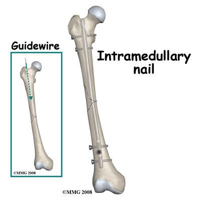
Thus the reason for treating a fractured femur by surgery is because the results of non-operative treatment are not consistently good. Surgery is done under general or spinal anesthetic. The bone is straightened and kept straight by traction. A small hole is then made at the top of the thigh bone and a thin wire is passed down inside the bone, crossing over the fracture and into the lower fragment. The inside of the bone may be reamed (cleared out) to ensure a snug fit. Next an Intramedullary Rod (IM Rod or IM Nail) is passed over the guide wire inside the femur and is secured with screws at either end.

With some fracture patterns or with open fractures, an external fixation device may be used. To apply an external fixator, the bone is straightened and large threaded pins are passed into the bone fragments above and below the fracture. These pins are attached to a rigid framework outside the thigh, which holds the fragments in position while the healing process takes place.

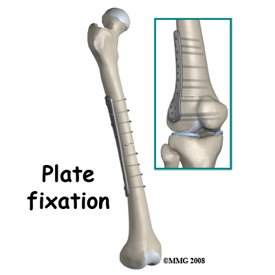
When the fracture extends far down the shaft towards the knee a plate may be used. The greatest advantage of operative treatment of a fractured femur is that it allows the patient freedom to move, to walk on crutches very soon after the surgery and to leave the hospital early. The bone is not healed by the surgery, but it is held still to improve the chances of healing. The quicker recovery of normal movement of the hip and knee prevents future problems of stiffness and weakness.

Implants are often removed after the bone is healed. The external fixators are always removed. Plates are also removed quite frequently as they may give the patient some symptoms and can often be felt through the skin. Removal of IM Rods is done only when they cause symptoms. The removal operation is relatively simple and recovery is usually quite rapid (six weeks) however, the bone may need some time to regain full strength after the hardware has been removed.
When there are no symptoms attributed to it, removal of the hardware is controversial. Some surgeons advocate it because the presence of a plate or a rod may weaken the bone long term; others leave the hardware in and point to the small but significant incidence of re-fracture in the three months after the implant is taken out.


